
Halaman ini berisi gambar-gambar ANATOMI dan FISIOLOGI serta PATOLOGI tubuh manusia. Anda bisa mendownload gambar-gambar tersebut dengan cara KLIK dahulu judul gambar dibawah ini lalu “KLIK KANAN” pada gambar tersebut dan pilih Save As —- arahkan ke direktori dimana Anda ingin menyimpan gambar tersebut.
.
NORMAL ANATOMY
.
A. HEAD and NECK
* Accessory Nerve – Schema * Action of Intrinsic Muscles of Larynx * Afferent Innervation of Mouth and Pharynx * Anterior and Posterior Chambers of Eye * Anterior Chamber Angle of Eye * Arteries and Veins of Hypothalamus and Hypophysis * Arteries and Veins of Orbit and Eyelids * Arteries of Brain – Frontal View and Section * Arteries of Brain – Inferior Views * Arteries of Brain – Lateral and Medial Views * Arteries of Nasal Cavity * Arteries of Oral and Pharyngeal Region * Arteries of Posterior Cranial Fossa * Arteries to Brain and Meningens * Arteries to Brain – Schema * Atlantooccipital Joint * Auditory Tube * Autonomic Innervation of Nasal Cavity * Autonomic Nerves in Head * Autonomic Nerves in Neck * Basal Ganglia * Bones of Cranial Base Superior View * Bony Framework of Head and Neck * Brainstem * Calvaria * Carotid Arteries * Cartilages of Larynx * Cerebellum * Cerebrum – Inferior View * Cerebrum – Lateral Views * Cerebrum – Medial Views * Cervical Plexus in Situ * Cervical Plexus – Schema * Cervical Vertebrae (1) * Cervical Vertebrae (2) * Cervical Vertebrae – Atlas and Axis * Ciliary Ganglion – Schema * Circle of Willis * Circulation of Cerebrospinal Fluid * Cranial Base – Inferior Views * Cranial Nerve Nuclei in Brainstem – Schema (1) * Cranial Nerve Nuclei in Brainstem – Schema (2) * Cranial Nerves (Motor and Sensory Distribution) – Schema * Cutaneous Nerves of Head and Neck * Deep Veins of Brain * External Craniocervical Ligaments * External Ear and Thympanic Cavity * External Nose * Extrinsic Eye Muscles * Eyeball * Eyelids * Facial Layers of Neck * Facial Nerves Branches and Parotid Gland * Facial Nerves – Schema * Fascia of Orbit and Eye * Fauces * Floor of Mouth * Foramina of Cranial Base – Superior View * Fourth Ventricle and Cerebellum * Glossopharyngeal Nerve – Schema * Hippocampus and Fornix * Hypoglossal Nerve – Schema * Hypothalamus and Hypophysis * Infrahyoid and Suprahyoid Muscles * Inspection of Larynx * Inspection of Oral Cavity * Intrinsic Arteries and Veins of Eye * Instrinsic Muscles of Larynx * Lacrimal Apparatus * Lateral Nasal Wall (1) * Lateral Nasal Wall (2) * Lens and Supporting Structure * Lymph Vessels and Nodes of Oral and Pharyngeal Region *Lymph Vessels and Nodes of Tongue and Pharynx * Mandible * Mandibular Nerves * Maxillary Arteries * Medial Nasal Wall * Meningeal Arteries * Meninges and Diploic Veins * Meninges and Superficial Cerebral Veins * Muscles Involved in Mastication (1) * Muscles Involved in Mastication (2) * Muscles of Facial Expression – Anterior View * Muscles of Neck – Anterior View * Muscles of Neck – Lateral View * Muscles of Pharynx – Lateral View * Muscles of Pharynx – Partially Opened – Posterior View * Muscles of Pharynx – Sagittal Section * Nerves of Larynx * Nerve of Nasal Cavity (1) * Nerve of Nasal Cavity (2) * Nerve of Oral and Pharyngeal Regions * Nerve of Orbit * Occulomotor, Trochlear and Abducens Nerves – Schema * Olfactory Nerve – Schema * Ophtalmic and Maxillary Nerve * Optic Nerve Visual Pathway – Schema * Osseus and Membranous Labyrinths * Osseus and Membranous Labyrinths (2) * Otic Ganglion – Schema * Paranasal Sinuses (1) * Paranasal Sinuses (2) * Paranasal Sinuses – Change with Age * Parathyroid Glands * Pathway of Sound Reception * Pharynx – Sagittal Section * Pterygopalatine and Submandibular Ganglia – Schema * Roof of Mouth * Salivary Glands * Scalene and Prevertebral Muscles * Skull of Newborn * Skull – Anterior View * Skull – Lateral View * Skull – Midsagittal Section * Subclavian Arteries * Subpendymal Veins of Brain * Superficial Arteries and Veins of Face and Scalp * Superficial Veins and Cutaneous Nerves of Neck * Taste Pathways -Schema * Teeth * Teeth (2) * Temporomandibular Joint * Thalamus * Thyroid Glands and Pharynx Posterior View * Thyroid Gland – Anterior View * Tongue (1) * Tongue (2) * Tongue and Mouth Section * Trigeminal Nerve – Schema *Tympanic Nerve * Vagus Nerve – Schema * Veins of Oral and Pharyngeal Regions * Veins of Cranial Posterior Fossa * Venous Sinuses of Duramater (1) * Venous Sinuses of Duramater (2) * Ventricle of Brain * Vestibulocochlear Nerve – Schema
.
B. BACK and SPINE
* Arteries of Spinal Cord – Intrinsic Distribution * Arteries of Spinal Cord – Schema * Autonomic Nervus System – General Topography * Autonomic Nervus System – Schema * Cholinergic and Adrenergic Synapses – Schema * Dermatomes * Lumbar Region of Back – Cross Section * Lumbar Vertebrae * Muscles of Back – Deep Layer * Muscles of Back – Intermediate Layers * Relation of Spinal Nerve Root to Vertebrae * Muscles of Back – Superficial Layers * Nerves of Back * Spinal Cord Cross Section – Fiber Tract * Spinal Cord In Situ * Spinal Membrane and Nerve Roots * Spinal Nerve Origin – Cross Section * Suboccipital Triangle * Thoracic Vertebrae * Typical Thoracic Spinal Nerve * Veins of Spinal Cord * Vertebral Column * Vertebral Ligaments – Lumbar Region * Vertebral Ligaments – Lumbosacral Region
.
C. THORAX
* Anterior Thoracic Wall (1) * Anterior Thoracic Wall (2) * Anterior Thoracic Wall – Internal View * Arteries of Esophagus * Arteries of Mammary Glands * Atria – Ventricles and Interventricular Septum * Autonomic Nerves in Thorax * Bony Framework of Thorax * Bronchial Arteries and Veins * Bronchopulmonary Segment (1) * Bronchopulmonary Segment (2) * Cervical Ribs and Related Anomalies * Conduction System of Heart * Coronary Arteries and Cardiac Veins * Coronary Arteries and Cardiac Veins – Variations * Coronary Arteries – Arteriographic Views * Costovertebral Joint * Diaphragm – Abdominal Surface * Diaphragm – Thoracic Surface * Esophagogastric Junction * Esophagus In Situ * Heart – Anterior Exposure * Heart – Basal and Diaphragmatic Surfaces * Heart In Situ * Innervation of Blood Vessels – Schema * Innervation of Heart – Schema * Innervation of Tracheobronchial – Schema * Intercostal Nerve and Arteries * Intrapulmonary Blood Circulation – Schema * Intrinsic Nerves and Variations in Nerves of Esophagus * Left Atrium and Ventricle * Lung In Situ – Anterior View * Lungs – Medial View * Lymph Vessels and Nodes of Esophagus * Lymph Vessels and Nodes of Lung * Lumph Vessels and Nodes of Mammary Gland * Mediastinum – Left Lateral View * Main Bronchi with Pulmonary Arteries and Veins In Situ * Mammary Glands * Mediastinum – Cross Section * Mediastinum – Right Lateral View * Muscles of Respiration * Musculature of Esophagus * Nerve of Esophagus * Nerve of Heart – Schema * Nomenclature of Bronchii – Schema * Pericardial Sac * Pharyngoesophageal Junction * Phrenic Nerve * Posterior and Lateral Thoracic Walls * Posterior Thoracic Wall * Prenatal and Postnatal Circulation * Pulmonary Arteries and Veins * Ribs and Sternocostal Joints * Right Atrium and Ventricle * Topography and Constrictions of Esophagus * Topography of Lung – Anterior View * Topography of Lung – Posterior View * Trachea and Major Bronchi * Valves of Heart (1) * Valves of Heart (2) * Veins of Esophagus
.
D. ABDOMEN
* Anterior Abdominal Wall – Deep Dissection * Anterior Abdominal Wall – Intermediate Dissection * Anterior Abdominal Wall – Superficial Dissection * Anterior Abdominal Wall – Internal View * Bony Framework of Abdomen * Arteries of Anterior Abdominal Wall * Femoral Sheath and Inguinal Canal * Inguinal and Femoral Region * Inguinal Region – Dissection * Inguinal Canal and Spermatic Cord * Nerve of Anterior Abdominal Wall * Posterolateral of Abdominal Wall * Rectus Sheath – Cross Section * Thoraco-Abdominal Nerve * Veins of Anterior Abdominal Wall
.
E. PELVIS and PERINEUM
* Anorectal Musculature * Arteries and Veins of Pelvic Organs – Female * Arteries and Veins of Pelvic – Female * Arteries and Veins of Pelvic – Male * Arteries and Veins of Perineum and Uterus * Arteries and Veins of Perineum – Male * Arteries and Veins of Testis * Arteries of Rectum and Anal Canal * Bones and Ligaments of Pelvis (1) * Bones and Ligaments of Pelvis (2) * Descent of Testis * Endopelvic Fascia and Spaces * External Anal Sphincter Muscle – Perineal View * Homologues of External Genitalia * Homologues of Internal Genitalia * Innervation of Female Reproductive Organs – Schema * Innervation of Male Reproductive Organs – Schema * Innervation of Urinary Bladder and Lower Ureter – Schema * Ischiorectal Fossa * Lymph Vessels and Nodes of Pelvis and Genitalia – Female * Lymph Vessels and Nodes of Pelvis and Genitalia – Male * Lymph Vessels and Nodes of Perineum – Male * Nerve of External Genitalia – Male * Nerve of Pelvic Viscera – Male * Nerve of Pelvic Viscera – Female * Nerve of Perineum and External Genitalia – Female * Neuropathways in Parturition * Ovary, Ova and Follicles * Pelvic Content – Female * Pelvic Content – Male * Pelvic Diaphragm – Female (1) * Pelvic Diaphragm – Male (2) * Pelvic Viscera and Perineum – Female * Pelvic Viscera and Perineum – Male * Pelvis Viscera – Female * Penis * Perineopelvic Space * Perineum (Superficial Dissection) – Female * Perineum and External Genitalia (Deeper Dissection) – Male * Perineum and External Genitalia (Superficial Dissection) – Male * Perineum and External Genitalia – Female * Perineum and Urogenital Diaphragm – Female * Prostate and Seminal Vesicle * Rectum and Anal Canal * Rectum In Situ – Female and Male * Scrotum and Contents * Sex Differences of Pelvis – Measurements * Testis, Epididymis and Ductus Deferens * Urethra – Female * Urethra – Male * Urinary Bladder – Female and Male * Urinary Bladder – Orientation and Supports * Urogenital Diaphragm – Male * Uterus and Adnexa * Uterus – Age Chanes and Muscle Pattern * Uterus – Vagina and Supporting Structures * Uterus – Variations in Position * Veins of Rectum and Anal Canal
.
F. UPPER LIMB
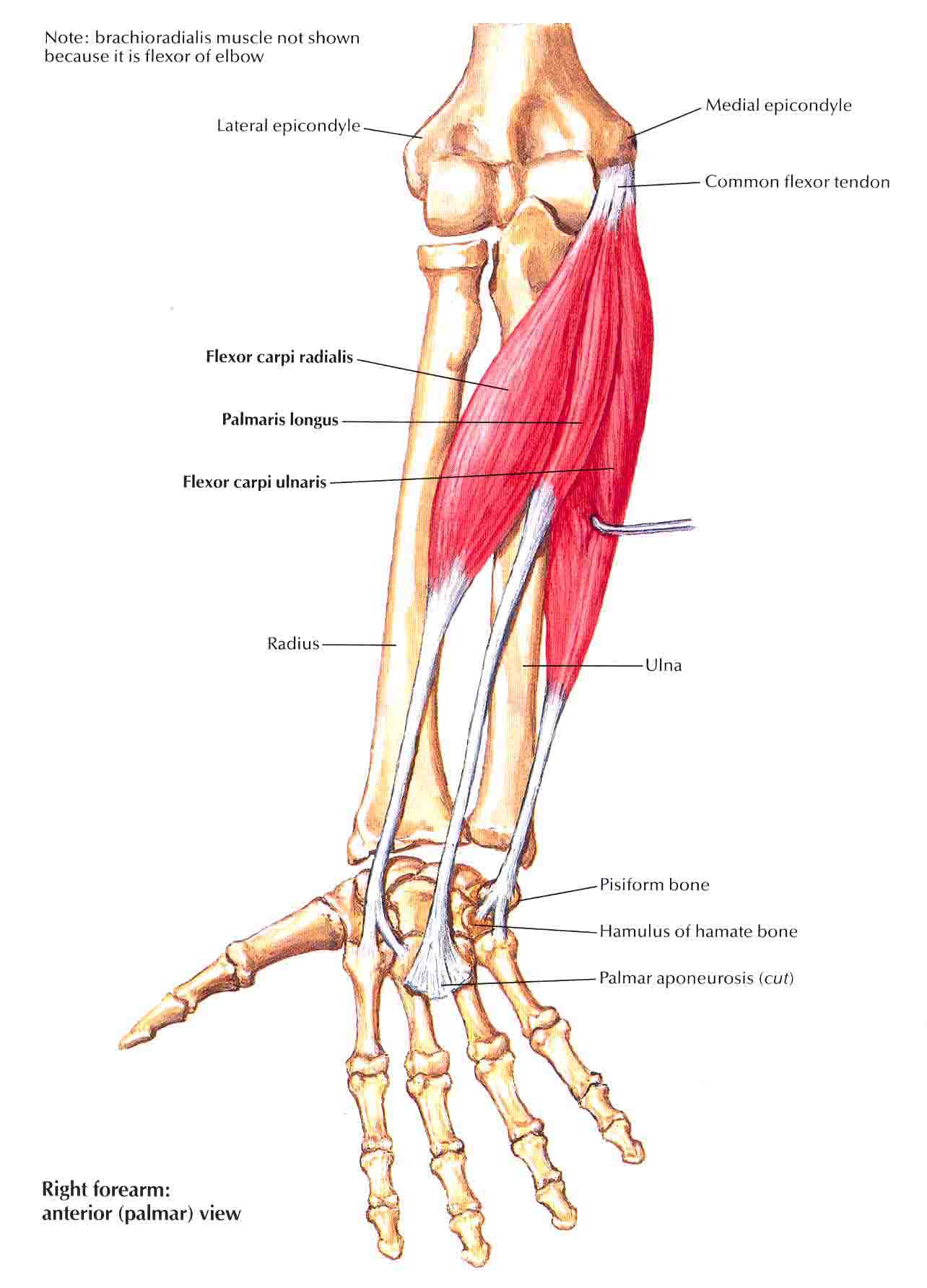
* Arm – Serial Cross Section * Arteries and Nerves of Hand – Palmar Views * Axilla (Dissection) – Anterior View * Axillary Artery and Anastomoses Around Scapula * Bones of Elbow * Bones of Forearm * Bones of Wrist and Hand * Bony Attachment of Muscles of Forearm – Anterior View * Bony Attachment of Muscles of Forearm – Posterior View * Brachial Artery and Anastomoses Around Elbow * Brachial Artery In Situ * Brachial Plexus – Schema * Bursae – Spaces and Tendon Sheaths of Hand * Carpal Bones * Clavicle and Sternoclavicular Joint * Flexor and Extensor Tendons in Finger * Flexor Tendons – Arteries and Nerves at Wrist * Forearm – Serial Cross Sections * Glenohumeral Joint * Humerus and Scapula – Anterior View * Humerus and Scapula – Posterior View * Individual Muscles of Forearm – Extensor of Wrist and Digits * Individual Muscles of Forearm – Flexors of Digits * Individual Muscles of Forearm – Flexors of Wrist * Individual Muscles of Forearm – Rotators of Radius * Intrinsic Muscles of Hand * Ligament of Elbow * Ligament of Wrist (1) * Ligament of Wrist (2) * Lumbrical Muscles and Bursae – Space and Sheath – Schema * Metacarpophalangeal and Interphalangeal Ligaments * Movement of Wrist * Muscle of Rotator Cuff * Muscle of Forearm (Deep Layer) – Anterior View * Muscle of Forearm (Deep Layer) – Posterior View * Muscle of Forearm (Intermediate Layer) – Anterior View * Muscle of Forearm (Superficial Layer) – Anterior View * Muscle of Forearm (Superficial Layer) – Posterior View * Muscles of Shoulder * Pectoral – Clavipectoral and Axillary Fasciae * Scapulohumeral Dissection * Wrist and Hand – Deeper Palmar Dissections * Wrist and Hand – Superficial Palmar Dissections * Wrist and Hand – Deeper Radial Dissections
.
G. LOWER LIMB
* Arteries and Nerves of Thigh – Anterior Views (1) * Arteries and Nerves of Thigh – Anterior Views (2) * Arteries and Nerves of Thigh – Posterior Views * Arteries of Femoral Head and Neck * Arteries of Thigh and Knee – Schema * Bones of Foot (1) * Bones of Foot (2) * Bony Attachments of Muscles of Hip and Thigh – Anterior View * Bony Attachments of Muscles of Hip and Thigh – Posterior View * Bony Attachments of Muscles of Leg * Calcaneus * Common Peroneal Nerve * Coxal Bone * Dermatome of Lower Limb * Dorsum of Foot – Deep Dissection * Femor * Femoral Nerve and Lateral Femoral Cutaneous Nerve * Hip Joint * Interosseous Muscles and Deep Arteries of Foot * Interosseous Muscles of Foot * Knee – Anterior View * Knee – Cruciate and Collateral Ligaments * Knee – Inferior View * Knee – Lateral and Medial Views * Knee – Posterior and Sagittal Views * Leg – Cross Section and Fascial Compartments * Ligament and Tendon of Ankle * Ligament and Tendons of Foot – Plantar View * Lumbar Plexus * Lumbosacral and Coccygeal Plexus * Lymph Vessels and Nodes of Lower Limb * Muscles of Dorsum of Foot – Superficial Dissection * Muscles of Hip and Thigh – Lateral View * Muscles of Hip and Thigh – Posterior View * Muscles of Leg (Deep Dissection) – Anterior View * Muscles of Leg (Deep Dissection) – Posterior View * Muscles of Leg (Intermediate Dissection) – Posterior View * Muscles of Leg (Superficial Dissection) – Anterior View * Muscles of Leg (Superficial Dissection) – Posterior View * Muscles of Leg – Lateral View * Muscles of Sole of Foot – First Layer * Muscles of Sole of Foot – Second Layer * Muscles of Sole of Foot – Third Layer * Nerves of Hip and Buttock * Obturator Nerve * Psoas and Iliacus Muscles * Sacral and Coccygeal Plexus * Sciatic Nerve and Posterior Femoral Cutaneous Nerve * Sole of Foot – Superficial Dissection * Superficial Nerves and Veins of Lower Limb – Anterior View * Superficial Nerves and Veins of Lower Limb – Posterior View * Tendon Sheath of Ankle * Thigh – Serial Cross Section * Tibia and Fibula (1) * Tibia and Fibula (2) * Tibial Nerve
.
PATHOLOGY BEDAH
* Appendicitis *Acute Appendicitis * Appendiceal Rupture * Appendicitis (2) * Appendicitis and Crohn Disease * Appendicitis Perforata (1) * Appendicitis Perforata (2) * Appendix Ruptured with Periappendicular Abscess * Appendix with Abscess *Abdominal and Thoracic Repair * Abdominal Aortic Aneurysm (1) * Abdominal Aortic Aneurysm (2) * Abdominal Mass * Abdominoplasty (1) * Abdominoplasty (2) * Aberrent of Biliary System * Anastomosis Leakage of Ileo-colica * Anatomy of a Four Year Old Child * Anatomy of Retroperitoneal Space (1) * Anatomy of Retroperitoneal Space (2) * Anatomy of the Kidney * Anterior and Posterior Spinal Fusion Procedure * Anterior L4-5 Interbody Fusion * Anterior Lumbar Fusion Using BAK Cages * Aorta to SMA Bypass * Bile Leak with ARDS * Bladder and Rectal Prolapsed * Bladder Augmentation with Ileal and Mitrofanoff Procedure * Bladder Removal-Ileal Loop and Urostomy bag * Bowel Injury and Perforation * Bowel Injury-Adhesions causing Obstruction of Arteries * Bowel Ischemia * Bowel Puncture during Laparoscopic * Bowel Resection with Wound Closure * Cecostomy * Cholecystectomy * Cholecystectomy Prosedure (1) * Cholecystectomy Procedure (2) * Cholecystectomy with Injury to the CBD * Cholecystitis with Subsequent Rupture * Chronic Open Abdominal Wound * Colchicine Toxicity * Colitis Ulcerative * Colon Cancer (1) * Colon Cancer (2) * Colon Lesion * Colon Polyps * Colonic Ischemic with Toxic Megacolon * Colonoscopy * Colonoscopy Procedure * Colonoscopy with Removal of Polyp * Colorectal Mass * Colostomy Irigation * Colostomy Pouch * Coronary Artery Blockage * Crohn Disease * Damaged Sigmoid Colon with Creation of Colostomy * Dangers of Hyposplenia * Diaphragmatic Hernia * Diarrhea Resulting from Loss of Terminal Ileum * Diastasis of Rectal Muscle with Bowel Adhesion * Diverticulitis * Diverticulitis-Ulcerative Colitis and Polyps * Double J Stent for Constricted Ureter * Drainage of Pelvic Abscess * Effects of Lower Spinal Cord Compression * Encephalopathy * Endoscopic in Massive Reflux and Lung Damage * Endoscopic Upper GI * Endotracheal Tube Insertion * Enterocutaneous Fistulas * Epidural Anesthesia * ERCP (1) * ERCP (2) * ERCP (3) * Erection Device Procedure * Esophageal Injury with Repair * Exploratory Laparotomy * Extensive Abdominoplasty * Failed Cholecystectomy with Leaking Cystic Duct * Female with Internal Body Organs * Femoral Nerve Injury * Function of Digestive System * Gall Stones (1) * Gall Stones (2) * Gallbladder * Gallbaldder Removed * Gas Exchange in the Placenta * Gastric Bypass * Gastric Volvulus * Gastroesophageal Reflux * Gastrostomy and Tracheostomy * Gastrostomy Tube (1) * Gastrostomy Tube (2) * Gastrostomy Tube (3) * Gastrostomy Tube (4) * Hartmann’s Procedure * Hemorrhaging Duodenal Ulcer * Hemorrhoid grade 3-4 * Hepaticojejunostomy * Hernias * Iatrogenic Injury to Iliac Artery after Trocar Placement * Iatrogenic to Iliac Artery during Lumbar Spine Surgery * Iatrogenic Ureter Injury causing Laparoscopic * Ileal Anastomosis * Ileocolic Anastomosis Procedure * Ileostomy * Improper of Gastrostomy Tube * Infection of the Small Bowel * Inflamation and Adhesion of the Pelvis * Inflamed Pancreas * Inflamed Retrocecal Appendix * Inguinal Hernia Repair * Inguinal Hernia Repair with Complication * Injuries of the Rib-Hip-Brain-Spleen-Lungs * Injury to the CBD (1) * Injury to the CBD (2) * Insertion of Trocar in Abdomen * Instrument of Lap-Chole * Intraoperative Uterine Perforation with Bowel Injury * Intraabdominal and Pelvic Adhesions * Intraoperative Bowel Perforation * Intraoperative Ureter Injuries * Ischemic Bowel with Resection * J-Pouch for Ileorectal Anastomosis * Kyphosis with Surgical Fusion * L5-S2 Disc Herniation with Bowel-Bladder Dysfunction * Laparoscopic * Laparoscopic Cholecystectomy with Bile Duct Injury * Laparoscopic Cholecystectomy with Conversion * Laparoscopic Colostomy * Laparoscopic Hernia Repair * Laparoscopic Inguinal Hernia Repair * Laparoscopic Insufflation * Laparoscopic Oophorectomy with Transection of Right Ureter * Laparoscopic Perforation of Bowel with Peritonitis * Laparoscopic to Remove Bowel Adhesion * Laparoscopic Ventral Hernia Repair * Laparoscopic with Bowel Perforation * Laparoscopic with Injury to the Iliac Vein * Laparoscopic vs Open Laparotomy * Laparotomy Incision Scar * Laparotomy with Bowel Resection * Lap-Chole vs Open Cholecystectomy * Liposuction of the Abdomen * Liver Damage * Liver Fibrosis * Lobectomy of the Lung * Lumbar Disc Herniation * Lumbar Fusion Procedure * Lumbar Sympathetic Injection with Bladder Neuropathy * Lymph Nodes of Abdominal * Lymphatic System * Lysing of Abdominal Adhesions * Mesenteric Ischemia * Mesenteric Torsion * Mesenteric Veins Trombosis * Metastasis of Colon Cancer to the Liver * Metastatic Testis Cancer * Methods of CBD * Mis-Injection into the Pancreatic Duct * Multiple Injuries * Nephrectomy (1) * Nephrectomy (2) * Nissen Funduplication (1) * Nissen Funduplication (2) * Pain Management * Pancreatic Cells * Penetrating Abdominal Injury * Percutaneous Endoscopic Gastrostomy (PEG TUBE) * Percutaneous Transhepatic Cholangiogram * Perforating Duodenal Ulcer * Perforating of Duodenal Ulcer with Peritonitis * Perforating of the Esophagus * Peritoneal Dialisis * Phimosis * Piece of Acromion * Pituitary Glands Deficiency * Placement of Greenfield Filter * Placement of Stent in Bile Duct * Plate and Screw for Fracture * Polyposis Coli * Post Accident Bowel Injuries with Repair * Post Accident Injuries to the Abdomen * Post Operative Adhesion * Post Operative Bowel Obstruction * Post Operative Cholecystectomy * Post Operative Cholecystectomy with Bile Leakage * Post Operative Obstruction of the Ureter * Posterior Abdominal Peritoneum * Pre Operative Bowel Obstruction * Prevention of Regurgitation and Aspiration * Progression of Abdominal Condition * Proper vs Improper Gastrostomy Tube * Prostate Cancer (1) * Prostate Cancer (2) * Prostate Cancer with Prostatectomy * Radiating Pain-Possible Areas * Recosntructive Billiary Surgery * Rectal Condition * Rectal Perforation * Recto- Sigmoidal Cancer * Recto-Vaginal Fistula * Recurrent Ventral Hernia * Removal of Gallbladder with Damage to the CBD * Removal of Polyps of the Colon * Removal of Portion of Esophagus and Stomach * Removed Portion of Terminal Ileum caused SBS * Repair of AAA * Repair of Bile Duct Injury * Repair of Rectal Perforation * Repair of the Diaphragm * Repair of Ureter with Psoas Hitch Procedure * Resection of Colorectal Mass * Resurrent Abscess with Open Drainage * Retained Sponge with Subsequent Infection * RES * Retroperitoneal Hematoma * Retroperitoneal Hemorrhage * Right Hemicolectomy Procedure * Roux-en-Y Construction * Roux-en-Y Construction of Gastrojejunostomy * Roux-en-Y Gastric Bypass (1) * Roux-en-Y Gastric Bypass (2) * Roux-en-Y Gastric Bypass (3) * Roux-en-Y Gastrojejunostomy * Roux-en-Y to Correct Billiary Tract Injury (1) * Roux-en-Y to Correct Billiary Tract Injury (2) * Ruptered Spleen * SBO with Regurgitation and Aspiration * Severe Diverticulitis and Phlegmon * Severe Gastroenteritis * Severe Pancreatitis * Sigmoid Colectomy with Transection of the Ureter * Sigmoidoscopy * SMA with Lack of Blood Supply to the Intestine * Small Bowel Obstruction(1) * Small Bowel Obstruction (2) * Spina Instability with Anterior Spinal Fusion * Splenectomy * Splenectomy with Incisional Hernia * Stapler for Resection of the Colon * Suprarenal Aortic Aneurysm * Surgical Removal of the Gallbladder * Surgical Repair of Hemorrhaging Duodenal Ulcer * Surgical Site Infection * Surgical Wound Healing * Surgical Wound Management * T6 Spinal Cord Injury * Temporary Colostomy for Infant * Torsion of Testis * Total Colectomy and Ileostomy * Tracheostomy-Gastrostomy and Jejunostomy * Tranfusion Syndrome * Traumatic Insertion of Trocar to the Small Bowel * Traumatic Rupture of Diaphragm * Traumatic to the Thoracic and Abdominal * Treatment of Hypertension * Trocar Insertion Adjacent to Adherent Bowel * Ulcerated Colon Umbilical Catheter in Newborn * Umbilical Catether in Newborn * Untreated Fecal Impaction * Upper GI Bleeding * Ureteral Calculus with Attempted Ureteroscopy * Vagus Nerve Injury with Gastroparesis * Vasculature of Large Intestine * Ventral Hernia Repair * Villous Adenoma in Colon * Volvulus of Small Bowel * VP Shunt (1) * VP Shunt (2) * VP Shunt (3) * Choledocolithiasis * Cholelithiasis * Hemorrhoid * Intraabdominal Disorder * Pre-hospital of Forearm Injury * Pre-hospital of Forearm Fracture * Pre-hospital of Hip Fracture * Pre-hospital of Leg Fracture * Pre-hospital of Fracture * Peptic Ulcer (1) * Peptic Ulcer (2) * Peritonitis * Rectal Prolapse * Staging of Colo-rectal Cancer * Visceral Trauma Associated Pelvic Fracture * Volvulus
16 Komentar
Comments RSS TrackBack Identifier URI


wah bagus sekali, terima kasih telah memasukan gambar-gambar ini ke web site. Tuhan memberkati pemilik address inii.
terima kasih atas kunjungannya dam doanya mas bambang…..semoga bermanfaat, amiinn
Mas Alex…..sip. semoga manfaat barokah
amiinnn terima kasih atas doanya
Terima Kasih Banyak atas File-file diatas, sangat bermanfaat. Tetapi ada beberapa File yang tidak bisa di akses, mohon diperbaiki dan kirim pemberitahuan ke Email saya rusmandeni@yahoo.co.id berikut ini adalah file yg tidak dapat di unduh;
Terima Kasih
terima kasih kembali, insyaAllah akan dicek nanti bila ada waktu
Yes! Finally someone writes about amoxicillin.
Hi there all, here every one is sharing such knowledge,
thus it’s good to read this web site, and I used to visit this webpage daily.
thank’s for your visiting
thank’s for your visiting…
perkenalkan.. saya Fachrisatul Masruroh I FAO PPDS bedah umum Unair.. senang sekali mengunjungi blog anda, terimakasih sdh menularkan banyak ilmu. semoga ilmu anda barokah dan bermanfaat untuk semua orang 🙂
amiiinnn…sama-sama dik…semoga bermanfaat bagi PPDS bedah umum lainnya
Terima kasih…ilmunya sangat bermanfaat…
muscle-up
ANATOMI-PATOLOGI ORGAN | Bedahunmuh’s Blog
Terima kasih sangat2 bermenafaat ilmunya.. mohon di download ya tuan.
ya sama-sama, terima kasih atas kunjungannya